

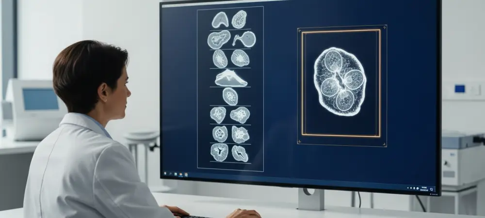
Modern pathology laboratories are currently navigating a monumental transition as the century-old practice of examining two-dimensional tissue slices evolves into a sophisticated era of three-dimensional digital analysis. This shift is not merely a change in perspective but a complete overhaul of how diagnostic data is harvested and interpreted by clinical experts. For generations, the gold standard relied on cutting biopsies into segments thinner than a human hair, a process that inherently ignores ninety-nine percent of the actual specimen. This massive sampling gap has historically posed a significant risk, as localized clusters of aggressive cancer cells can easily hide within the unexamined gaps between those physical slices. To address this structural vulnerability, researchers have introduced advanced imaging systems that provide a holistic view of the tissue. However, this wealth of data has created a secondary crisis: a volume of information so vast that human observation alone cannot possibly keep pace with the demands of a modern hospital.
Addressing the Data Deluge: The Rise of Volumetric Imaging
The implementation of open-top light-sheet microscopy has fundamentally changed the landscape by allowing clinicians to capture entire tissue volumes without the destructive process of physical sectioning. This optical innovation creates a continuous digital map of every cell, gland, and vascular structure within a specimen, offering a level of transparency previously thought impossible in routine diagnostics. Despite these benefits, the sheer scale of the output is staggering, with a single biopsy potentially generating thousands of high-resolution digital layers. If a pathologist were to manually review each of these images, the diagnostic process for a single patient would extend from minutes into hours or even days. This bottleneck threatened to stall the adoption of 3D imaging in clinical settings until the development of specialized artificial intelligence frameworks designed to process these massive datasets. These systems act as a critical bridge, turning raw volumetric data into actionable insights for the medical team.
The TRICARE system was specifically engineered to alleviate this pressure by functioning as an intelligent triage assistant that evaluates every cross-section within a 3D volume. Rather than presenting a chaotic mountain of images, the AI assigns a precise risk score to each individual layer based on detected morphological anomalies. By identifying and highlighting the most suspicious regions, the software automatically reorganizes the digital stack, pushing the highest-risk data points to the front of the review queue for the pathologist. This prioritized workflow ensures that the clinician’s attention is immediately directed toward the most critical biological indicators, effectively filtering out thousands of healthy or redundant frames. This method of selective focus does not just save time; it maximizes the utility of human expertise by ensuring that every minute spent by a doctor is dedicated to evaluating areas with the highest probability of containing life-threatening pathology or early-stage indicators of disease.
Context-Aware Intelligence: Deep Learning Beyond Snapshots
Traditional machine learning models in pathology often struggle because they were designed to analyze flat, isolated snapshots that lack the depth and continuity of actual biological tissue. The TRICARE framework overcomes this limitation by employing context-aware deep-learning algorithms that recognize how structures like glands or tumor nests exist in three dimensions. By analyzing an image in relation to the layers immediately above and below it, the system gains a holistic understanding of spatial continuity that mimics the way human vision perceives physical objects. This architectural approach allows the AI to track the winding paths of complex anatomical features, ensuring that fragmented views of a single structure are correctly identified as part of a larger whole. Such a capability is essential for accurate diagnostics, as many diseases are characterized not just by the appearance of individual cells, but by the abnormal growth patterns and architectural distortions that only become apparent when viewed across multiple planes of volume. One of the most significant advantages of this spatial awareness is its ability to distinguish between actual pathological changes and common imaging artifacts that frequently plague digital pathology. In the preparation and scanning of biological specimens, it is common to encounter tissue folds, staining variations, or air bubbles that can mimic the appearance of malignancy to an untrained algorithm. However, because TRICARE evaluates the three-dimensional consistency of these features, it can quickly identify if an anomaly is a physical defect in the sample or a genuine cellular mutation. Artifacts typically lack the logical spatial structure found in biological growths, appearing and disappearing abruptly between layers. By filtering out these false positives, the system provides a much cleaner dataset for the pathologist to review, significantly reducing the cognitive load associated with sorting through technical noise. This refined accuracy strengthens the partnership between the machine and the clinician, building trust in the AI’s ability to highlight only the most relevant findings.
Clinical Validation: Success in High-Stakes Diagnostics
The practical efficacy of this AI triage system has been rigorously demonstrated through validation studies focusing on complex medical conditions such as prostate cancer and Barrett’s esophagus. In prostate cancer diagnostics, identifying the highest-grade features of a tumor is essential for determining whether a patient requires aggressive surgery or active surveillance. The AI successfully navigated 3D volumes of prostate biopsies to pinpoint specific regions with high-grade patterns, often finding aggressive cells that would have been missed by standard 2D sampling techniques. Similarly, for patients with Barrett’s esophagus, the system proved adept at flagging rare precancerous changes that are frequently distributed sporadically across a wide area of tissue. By providing a comprehensive review of the entire specimen volume, the TRICARE framework ensured that these subtle markers were not overlooked, directly influencing the accuracy of the final diagnosis and the subsequent treatment plan for the patients involved in the study.
A central philosophy behind the integration of this technology is the maintenance of human oversight, ensuring the AI serves as a support tool rather than an autonomous decision-maker. The system does not issue a definitive diagnosis; rather, it functions as a high-speed lens that brings the most important features into sharp focus for the medical professional. This human-centric design is crucial for clinical acceptance, as it allows pathologists to apply their deep knowledge of patient history and clinical nuance to the data highlighted by the algorithm. The transparency of the triage process means that every risk score and highlighted layer can be independently verified by the doctor, maintaining a clear chain of accountability. This approach preserves the traditional diagnostic relationship while augmenting it with computational power, allowing for a more thorough examination of patient tissue without sacrificing the critical judgment that only a seasoned medical expert can provide in complex or borderline cases.
Operational Evolution: Infrastructure and Strategic Implementation
The successful adoption of 3D pathology required healthcare institutions to modernize their digital infrastructure to accommodate the storage and processing of massive volumetric files. Hospitals that integrated high-performance computing clusters and robust data management systems found they could run deep-learning models in real-time, providing immediate triage results as soon as a scan was completed. Pathologists shifted their focus toward reviewing high-risk segments identified by the system, which optimized the workflow and reduced the time spent on routine negative cases. Medical boards established clear guidelines for the use of AI in diagnostics, ensuring that software updates were validated against diverse patient populations to maintain consistency across different demographic groups and scanner types. These strategic investments turned 3D imaging from a niche research application into a central component of clinical practice, providing a more comprehensive understanding of disease while simultaneously increasing the throughput of busy pathology departments.
Moving forward, the focus moved toward expanding the application of these triage frameworks into other specialized areas of diagnostic medicine, including neuropathology and dermatopathology. Leaders in the field emphasized the importance of standardized protocols for data sharing and model training to ensure that the AI remains effective as imaging hardware continues to improve. By fostering a collaborative environment between software developers and clinical practitioners, the medical community ensured that the evolution of 3D pathology remained focused on improving patient outcomes through greater diagnostic precision. The integration of these tools demonstrated that when technology is used to handle the burden of data processing, clinicians are free to concentrate on the complex interpretive tasks that define high-quality healthcare. Ultimately, the lessons learned from the deployment of TRICARE paved the way for a more resilient and data-driven diagnostic landscape, where the risk of missing a life-altering diagnosis due to sampling limitations was significantly diminished.